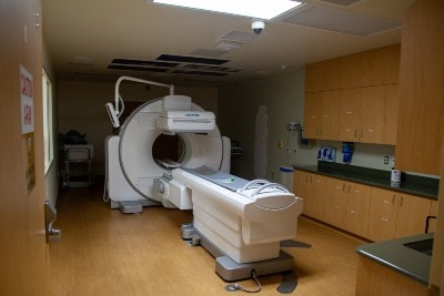
Nuclear Medicine
 The Torrance Memorial Radiology Department is at the forefront of nuclear medicine, technologies specialize in the imaging of an organ’s metabolic functions. This facilitates diagnosis of certain diseases, as well as identifying various medical conditions.
Nuclear imaging tests look for the biological changes in the body that indicate the presence of disease rather than changes in anatomy. Because of this basic difference, nuclear medicine imaging procedures can often identify abnormalities very early, sometimes before medical problems are apparent with other diagnostic tests.
Types of Nuclear Medicine Exams
Common conditions for which nuclear medicine imaging tests are used include:
- Visualize blood flow to the heart
- Bone scans for fractures and infections
- Lung scans for blood clots or pulmonary function
- Liver, kidney and gallbladder scans to diagnose abnormal function or blockages.
- Measure thyroid function
- Determine whether cancer is present or has spread in certain parts of the body
- Investigate abnormalities in the brain, such as seizures or memory loss
What to Expect During a Nuclear Medicine Exam
Nuclear medicine exams are safe, painless and usually well tolerated. Nuclear imaging uses a pharmaceutical drug that is attached to a small amount of radioactive material, called a radioisotope. This combination is called a radiopharmaceutical or radiotracer. Different radiotracers are available to study different parts of the body.
Radiotracers are introduced into the patient's body by injection, swallowing, or inhalation. After the radiotracer is administered, imaging will be done either immediately, a few hours later, or even several days after administration. Imaging times generally range from 20 to 45 minutes.
Gamma cameras produce nuclear medicine images. The gamma camera does not make any noise, nor does it transmit any radiation to the patient. Instead, the camera detects radiation that is being emitted from the patient's body.
Depending on the kind of pictures needed, the gamma cameras will operate in a stationary mode, move across the body, or rotate around the body. Sophisticated computer software is then used to process the images. A radiologist will interpret the images and forward a report to the referring physician. The report is typically available within 48 hours and can be discussed with you by your physician.
Nuclear Medicine Studies & Radiation
The Torrance Memorial Radiology Department is committed to performing high quality imaging studies while adhering to principles of radiation safety. Nuclear medicine technologists use the ALARA Principle (As Low As Reasonably Achievable) to carefully select the amount of radiotracer that will provide an accurate test with the least amount of radiation exposure to the patient.
After most nuclear medicine procedures it is generally advised to drink a lot of fluid and urinate as frequently as possible, as this helps to flush the remaining radioactivity out of the body. Your nuclear medicine technologist will advise you on the length of time you will need to do this.

Change Your Location


