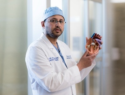
Cardiac Imaging
 At Torrance Memorial, we are proud to offer the newest and most advanced diagnostic technology to identify heart disease at its earlier and most treatable stages. With our state-of-the-art imaging tools we can discover problems that just a few years ago were undetectable using conventional methods of diagnosis.
The Torrance Memorial Cardiovascular Imaging program offers the full array of imaging technology to diagnose heart disease, plan optimal care for patients, and ensure that treatment is complete. We also use these diagnostic studies for pre-surgical planning to minimize complications during many types of catheter-based procedures and heart surgery.
Cardiac Computed Tomography (CT)
A cardiac CT scan measures the coronary artery calcium "score." This is a non-invasive way of obtaining information about the location and extent of calcified plaque in the coronary arteries, the vessels that supply blood to the heart muscle. Plaque is a build-up of fat and other substances, including calcium, which can, over time, narrow the arteries or even close off blood flow to the heart. The result may be painful angina in the chest or a heart attack. Because calcium is a marker of coronary artery disease, the amount of calcium detected on a cardiac CT scan is a helpful diagnostic marker.
Coronary Computed Tomography Angiography (CCTA)
A computerized tomography coronary angiogram (CCTA) is a non-invasive way to examine the health of the arteries that supply your heart muscle with blood. Taking just seconds, a CT angiogram involves injection of an IV contrast dye that helps produce highly detailed images of your heart and blood vessels. Often this test eliminates the need for a coronary angiogram, a more invasive and lengthy procedure which requires sedation, a catheter inserted into your groin, and a longer period of recovery. At Torrance Memorial, we have dedicated radiologists with specialized training and certification to interpret this type of CT scan.
Cardiac Magnetic Resonance Imaging (MRI)
Cardiac MRI (CMRI) is a noninvasive, radiation-free diagnostic test that allows doctors to obtain detailed images of the heart and adjacent structures. Cardiac MRI is helpful in diagnosing heart disease, heart attack, scarring, tumors and valvular diseases. Cardiac MRI can also be used to study abnormalities of the blood vessels arising from the heart and to measure blood flow through the aorta and aortic valve.
Cardiac Positron Emission Tomography (PET)
A cardiac PET study evaluates blood flow to the heart and is also used to evaluate damage to the heart muscle after a heart attack. A small amount of radioactive tracer is injected into a vein. A special camera, called a gamma camera, finds the radiation and uses it to produce computer images of the heart. This test can help evaluate whether there is enough blood flow to the heart during activity.
Echocardiogram (ECHO)
An echocardiogram is a noninvasive procedure used to assess the heart's function and structures. During the procedure, a transducer, similar to a microphone, sends out ultrasonic sound waves at a frequency too high to be heard. When the transducer is placed on the chest at certain locations and angles, the ultrasonic sound waves move through the skin and other body tissues to the heart tissues, where the waves bounce or "echo" off of the heart structures. These sound waves are sent to a computer that can create moving images of the heart walls and valves.
Learn More
Nuclear Cardiology
A nuclear stress test lets doctors see pictures of your heart while you are resting and shortly after you have exercised. The test provides information about the size of the heart's chambers, how well the heart is pumping blood, and whether the heart has any damaged or dead muscle. Nuclear stress tests can also give doctors information about your arteries and whether they are narrowed or blocked because of coronary artery disease.
How Will You Learn About Your Results?
The technologist will not give you the test results directly, as the images still need to be reviewed by a radiologist. After reviewing the study, the radiologist will send an official report to your physician, who can then discuss the results with you.

